
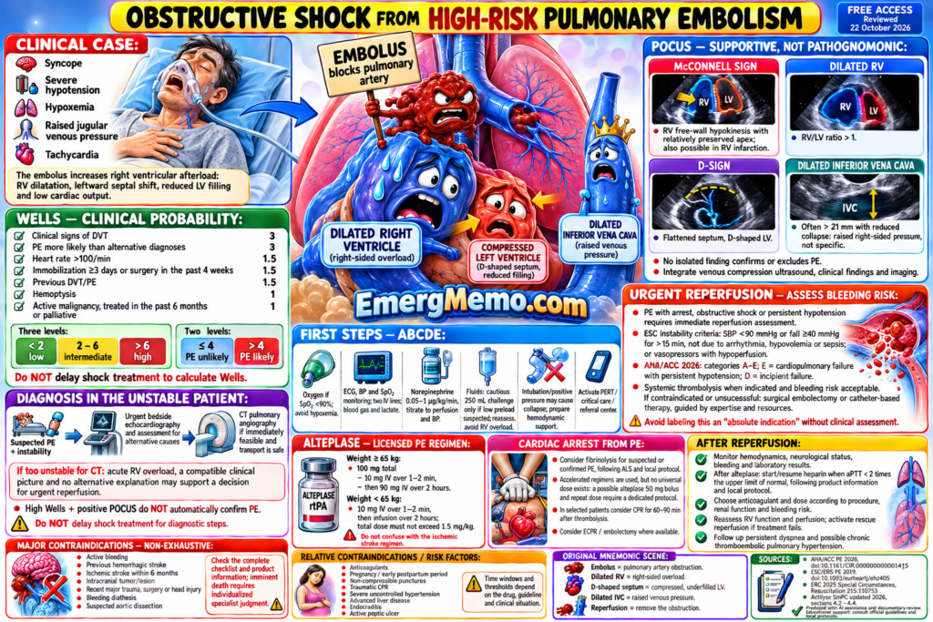
Free access — complete English version. Reworked from the original, with a 3D mnemonic scene and clinical review on 22 October 2026. Open the image to zoom.
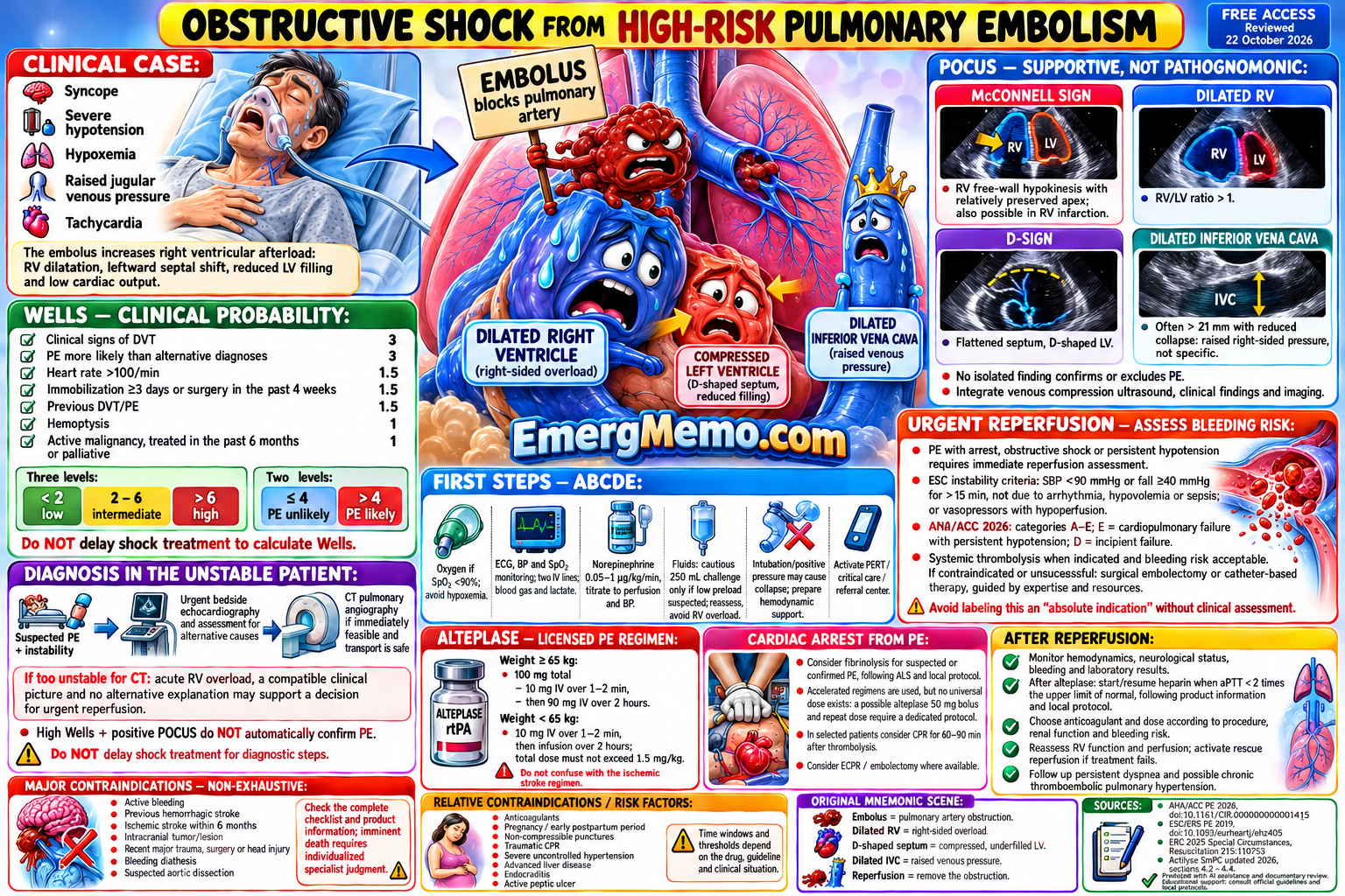
Complete readable text
OBSTRUCTIVE SHOCK FROM HIGH-RISK PULMONARY EMBOLISM
FREE ACCESS • Reviewed 22 October 2026
CLINICAL CASE:
Syncope • Severe hypotension • Hypoxemia • Raised jugular venous pressure • Tachycardia.
The embolus increases right ventricular afterload: RV dilatation, leftward septal shift, reduced LV filling and low cardiac output.
WELLS — CLINICAL PROBABILITY:
Clinical signs of DVT: 3
PE more likely than alternative diagnoses: 3
Heart rate >100/min: 1.5
Immobilization ≥3 days or surgery in the past 4 weeks: 1.5
Previous DVT/PE: 1.5
Hemoptysis: 1
Active malignancy, treated in the past 6 months or palliative: 1
Three levels: <2 low; 2–6 intermediate; >6 high.
Two levels: ≤4 PE unlikely; >4 PE likely.
Do NOT delay shock treatment to calculate Wells.
DIAGNOSIS IN THE UNSTABLE PATIENT:
Suspected PE + instability → urgent bedside echocardiography and assessment for alternative causes → CT pulmonary angiography if immediately feasible and transport is safe.
If too unstable for CT: acute RV overload, a compatible clinical picture and no alternative explanation may support a decision for urgent reperfusion.
High Wells + positive POCUS do NOT automatically confirm PE.
POCUS — SUPPORTIVE, NOT PATHOGNOMONIC:
• McConnell sign: RV free-wall hypokinesis with relatively preserved apex; also possible in RV infarction
• Dilated RV: RV/LV ratio >1
• D-sign: flattened septum, D-shaped LV
• Dilated inferior vena cava, often >21 mm with reduced collapse: raised right-sided pressure, not specific
• No isolated finding confirms or excludes PE
• Integrate venous compression ultrasound, clinical findings and imaging
FIRST STEPS — ABCDE:
• Oxygen if SpO₂ <90%; avoid hypoxemia
• ECG, BP and SpO₂ monitoring; two IV lines; blood gas and lactate
• Norepinephrine 0.05–1 µg/kg/min, titrate to perfusion and BP
• Fluids: cautious 250 mL challenge only if low preload suspected; reassess, avoid RV overload
• Intubation/positive pressure may cause collapse; prepare hemodynamic support
• Activate PERT / critical care / referral center
URGENT REPERFUSION — ASSESS BLEEDING RISK:
PE with arrest, obstructive shock or persistent hypotension requires immediate reperfusion assessment.
ESC instability criteria: SBP <90 mmHg or fall ≥40 mmHg for >15 min, not due to arrhythmia, hypovolemia or sepsis; or vasopressors with hypoperfusion.
AHA/ACC 2026: categories A–E; E = cardiopulmonary failure with persistent hypotension; D = incipient failure.
Systemic thrombolysis when indicated and bleeding risk acceptable. If contraindicated or unsuccessful: surgical embolectomy or catheter-based therapy, guided by expertise and resources.
Avoid labeling this an “absolute indication” without clinical assessment.
ALTEPLASE — LICENSED PE REGIMEN:
Weight ≥65 kg: 100 mg total — 10 mg IV over 1–2 min, then 90 mg IV over 2 hours.
Weight <65 kg: 10 mg IV over 1–2 min, then infusion over 2 hours; total dose must not exceed 1.5 mg/kg.
Do not confuse with the ischemic stroke regimen.
CARDIAC ARREST FROM PE:
Consider fibrinolysis for suspected or confirmed PE, following ALS and local protocol.
Accelerated regimens are used, but no universal dose exists: a possible alteplase 50 mg bolus and repeat dose require a dedicated protocol.
In selected patients consider CPR for 60–90 min after thrombolysis. Consider ECPR / embolectomy where available.
MAJOR CONTRAINDICATIONS — NON-EXHAUSTIVE:
Active bleeding; previous hemorrhagic stroke; ischemic stroke within 6 months; intracranial tumor/lesion; recent major trauma, surgery or head injury; bleeding diathesis; suspected aortic dissection.
Check the complete checklist and product information; imminent death requires individualized specialist judgment.
RELATIVE CONTRAINDICATIONS / RISK FACTORS:
Anticoagulants; pregnancy / early postpartum period; non-compressible punctures; traumatic CPR; severe uncontrolled hypertension; advanced liver disease; endocarditis; active peptic ulcer.
Time windows and thresholds depend on the drug, guideline and clinical situation.
AFTER REPERFUSION:
Monitor hemodynamics, neurological status, bleeding and laboratory results.
After alteplase: start/resume heparin when aPTT <2 times the upper limit of normal, following product information and local protocol.
Choose anticoagulant and dose according to procedure, renal function and bleeding risk.
Reassess RV function and perfusion; activate rescue reperfusion if treatment fails.
Follow up persistent dyspnea and possible chronic thromboembolic pulmonary hypertension.
ORIGINAL MNEMONIC SCENE:
Embolus = pulmonary artery obstruction.
Dilated RV = right-sided overload.
D-shaped septum = compressed, underfilled LV.
Dilated IVC = raised venous pressure.
Reperfusion = remove the obstruction.
References
- AHA/ACC Pulmonary Embolism 2026
- ESC/ERS Pulmonary Embolism 2019
- ERC 2025 — Special Circumstances
- Actilyse — SmPC / RCP, 2026
Produced with AI assistance and documentary review. Educational support for professionals: consult official guidelines, product information and local protocols.